Eye Floaters

Floater: A blurry spot that seems to drift in front of the eyes but does not block vision. The blur is the result of debris from the vitreous of the eye casting a shadow on the retina. The spot is the image formed by a deposit of protein drifting about in the vitreous, the clear jelly-like substance that fills the middle of the eye.

Floaters are often described by patients as spots, strands, or little flies.

Floaters are usually benign (not serious). They can, however, occasionally result from a separation of the vitreous gel from the retina. This condition is called a posterior vitreous detachment (PVD). Although a PVD occurs commonly, there are no retinal tears associated with the condition most of the time.
No treatments are available for floaters although they tend to become less annoying over time. Remember also that floaters are usually not associated with serious eye problems.
Permanent or recurring white or black spots in the same area of your field of vision may, however, be an early warning sign of cataracts or another serious eye problem. If you experience a shadow or curtain that affects any part of your vision, this can indicate that a retinal tear has occurred and has progressed to a detached retina. In this situation, you should immediately consult an ophthalmologist since time is of the essence.
Read more »
These icons link to social bookmarking sites where readers can share and discover new web pages.
  • Digg
  • Sphinn
  • del.icio.us
  • Facebook
  • Mixx
  • Google
  • Furl
  • Reddit
  • Spurl
  • StumbleUpon
  • Technorati

Visual acuity testing

The visual acuity score compares your distance vision with that of people who have normal vision, using an eye chart. Each eye's score is expressed as two numbers, such as 20/20 (6/6) or 20/100 (6/30). The first number is the distance you stand from the chart, usually 20 ft (6 m) when using a typical wall chart. The second number is the distance from which people with normal eyesight can read the same line on the eye chart. 


20/20 (6/6) vision is considered normal. A person with 20/20 vision can see at 20 ft (6 m) what people with normal vision can see at this distance.
  • When the second number is smaller than the first number, the person's vision is better than normal. For instance, a person with 20/10 (6/3) vision can see from 20 ft (6 m) what people with normal vision can see from 10 ft (3 m).
  • When the second number is larger than the first number, the person's distance vision is worse than normal.
  • A person with 20/200 (6/60) vision or less in his or her best eye when wearing corrective lenses is considered legally blind.
For near vision, 14/14 (35/35) is normal, with 14 in. (36 cm) being the normal distance for reading. If the second number is greater than 14 (14/20, for example, or 35/50), it means that you have reduced near vision. You have to be 14 in. (36 cm) away to read print that people with normal near vision can read from 20 in. (51 cm).
A Jaeger (J) number is another way to rate your near vision. The J number relates to the size of text you could read on the Jaeger chart. The J number goes up as the print size of the text you read goes up. The higher the J number, the worse your near vision. The number can range from J1 to J16. For example:
  • J1 means that you could read the smallest text on the chart and that you have 20/15 vision.
  • J2 means the line of text you were able to read had larger print than J1, and your vision is 20/20.
  • J3 means the line of text you were able to read had larger print than J1 and J2, and your vision is 20/40.
Visual acuity tests usually take about 5 to 10 minutes
Read more »
These icons link to social bookmarking sites where readers can share and discover new web pages.
  • Digg
  • Sphinn
  • del.icio.us
  • Facebook
  • Mixx
  • Google
  • Furl
  • Reddit
  • Spurl
  • StumbleUpon
  • Technorati

Vision Tests-Refraction

Refraction

Refraction is a test that measures the eye's need for a corrective lens (refractive error). For this test, you will be asked to describe the effects of looking at an eye chart through various corrective lenses.
Your health professional may use eyedrops to widen (dilate) your pupils before you start this test. The eyedrops take about 15 to 20 minutes to dilate the pupil fully.
Using a retinoscope, your health professional may shine light into your eyes. A series of trial lenses will be placed in front of your eyes and adjusted until the light rays are properly focused on your retina. Testing one eye at a time, the health professional will ask you to compare the effects of two lenses (first one lens, then the other). You should state which lens of each pair gives you better vision. The health professional will continue to test your eyes with different lenses until it is determined which lenses correct your vision the best. 

Visual field tests

Visual field tests are used to check for gaps in your range of vision. They can help detect eye diseases or nervous system problems that limit your ability to see objects clearly in the entire visual field or in one part of it. Several tests are commonly done to evaluate a person's visual field.
  • The confrontation test. Your doctor will sit or stand 2 ft (0.6 m) to 3 ft (1 m) in front of you. You cover one eye while fixing your gaze on his or her nose. He or she slowly moves a finger or hand from the outer edge of your visual field toward the center and from the center toward the edge through all areas of your visual field. You will focus your eye on your doctor's nose and signal when you first see his or her finger or hand. The test is then repeated for the other eye.
  • The Amsler grid test checks for macular degeneration, a disease that causes loss of vision in the center of your visual field. The test uses a 4 in. (10 cm) square chart with straight lines that form boxes. The grid has a black dot at the center. The chart is held about 14 in. (36 cm) from your face. You will cover one eye while focusing your other eye on the black dot. The test is then repeated on the other eye. Tell your health professional if:
    • You cannot see the black dot.
    • You see a blank or dark spot (other than the center dot).
    • The lines in the grid look wavy, blurred, or curved instead of straight. You will be asked to point to the specific abnormal area of the grid.
  • Perimetry testing uses a machine that flashes lights randomly at various points in the visual field. You look inside a bowl-shaped instrument called a perimeter. While you stare at the center, lights will flash, and you press a button each time you see a flash. A computer records the location of each flash and whether you pressed the button when the light flashed in that location. At the end of the test, a printout shows any areas of your visual field where you did not see the flashes of light. In an alternative manual perimetry test, your health professional moves a light target and notes your visual field on paper.
  • The tangent screen test uses a black screen with concentric circles and lines leading out from a center point (like a bull's-eye). Sitting 3 ft (1 m) to 6 ft (2 m) away from the screen, you cover one eye while fixing your gaze on a target point marked on the screen. Test objects of various sizes at the tip of a wand are then moved inward from the outer edge of the screen toward the center. You will signal when you can see the object, and that point is then marked on the screen. The points on the screen where you see the objects are connected to provide an outline of your visual field. The test is then repeated for the other eye. An alternative manual tangent screen test uses a white object against a black background. If you wear glasses, you will keep them on for this test.
Color vision tests check your ability to distinguish colors. In the most commonly used color vision test, you look for different colored numbers or symbols hidden in varying backgrounds of colored dots.
First, you are shown sample patterns and told what symbols and numbers you can expect to see. You then sit at a table and cover one eye. The health professional holds the color test patterns about 14 in. (36 cm) away from you. Some patterns are more difficult to pick out than others. As the health professional holds up a pattern, you will identify the number or symbol you see and trace it using a pointer. Some patterns may not have a number or symbol. The test is then repeated with the other eye.
Read more »
These icons link to social bookmarking sites where readers can share and discover new web pages.
  • Digg
  • Sphinn
  • del.icio.us
  • Facebook
  • Mixx
  • Google
  • Furl
  • Reddit
  • Spurl
  • StumbleUpon
  • Technorati

Vision Tests-2

How It Is Done

Visual acuity testing

Visual acuity tests are used to evaluate eyesight. Several types of visual acuity tests may be used.

  • The Snellen test checks your ability to see at distances. It uses a wall chart that has several rows of letters. The letters on the top row are the largest; those on the bottom row are the smallest.
    • You will stand or sit 20 ft (6 m) from the chart and be asked to cover one eye and then read the smallest row of letters you can see on the chart. If you are unable to cover your eye, an eye patch will be placed over your eye.
    • Each eye is tested separately. You may be given a different chart or asked to read a row backward to make sure that you did not memorize the sequence of letters from the previous test.
    • If you wear glasses or contacts, you may be asked to repeat the test on each eye while wearing them.
    • Let your health professional know if you have trouble reading the letters on one side of the row, or if some letters disappear while you are looking at other letters. You may have a visual field problem, and visual field tests may be needed.
  • The E chart tests the vision of children and people who cannot read. The E chart is similar to the Snellen chart in that there are several rows, but all of the rows contain only the letter E in different positions. The top row is the largest and the bottom row of Es is the smallest. You will be asked to point in the same direction as the lines of the E. Similar charts use the letter C or pictures. These charts are also available in a handheld card.
  • The Near test uses a small card (Jaeger chart) containing a few short lines or paragraphs of printed text to test your near vision. The size of the print gradually gets smaller. You will be asked to hold the card about 14 in. (36 cm) from your face and read aloud the paragraph containing the smallest print you can comfortably read. Both eyes are tested together, with and without corrective lenses. This test is routinely done after age 40, because near vision tends to decline as you age (presbyopia).
If you cannot read any of the letters or print on these charts because of poor vision, your visual acuity will be tested by other techniques, such as counting fingers, detecting hand movements, or distinguishing the direction or perception of light sources (such as room light or a penlight held up close to the face).
Visual acuity tests usually take about 5 to 10 minutes. They may be performed by a nurse, a medical assistant, an ophthalmologist, an optometrist, a teacher, or some other trained person. Testing may be done at a doctor's office, school, workplace, health fair, or elsewhere.
Read more »
These icons link to social bookmarking sites where readers can share and discover new web pages.
  • Digg
  • Sphinn
  • del.icio.us
  • Facebook
  • Mixx
  • Google
  • Furl
  • Reddit
  • Spurl
  • StumbleUpon
  • Technorati

Vision Tests-1

Vision tests check many different functions of the eye. The tests measure your ability to see details at near and far distances, check for gaps or defects in your field of vision, and evaluate your ability to see different colors.

  • Visual acuity tests are the most common tests used to evaluate eyesight. They measure the eye's ability to see details at near and far distances. The tests usually involve reading letters or looking at symbols of different sizes on an eye chart. Usually, each eye is tested by itself. And then both eyes may be tested together, with and without corrective lenses (if you wear them). Several types of visual acuity tests may be used.
  • Refraction is a test that measures the eyes' need for corrective lenses (refractive error). It is usually done after a visual acuity test. Refractive errors, such as nearsightedness or farsightedness, occur when light rays entering the eye can't focus exactly on the nerve layer (retina) at the back of the eye. This causes blurred vision. Refraction is done as a routine part of an eye examination for people who already wear glasses or contact lenses, but it will also be done if the results of the other visual acuity tests show that your eyesight is below normal and can be corrected by glasses.
  • Visual field tests are used to check for gaps in your side (peripheral) vision. Your complete visual field is the entire area seen when your gaze is fixed in one direction. The complete visual field is seen by both eyes at the same time, and it includes the central visual field-which detects the highest degree of detail-and the peripheral visual fields.
  • Color vision tests check your ability to distinguish colors. It is used to screen for color blindness in people with suspected retinal or optic nerve disease or who have a family history of color blindness. The color vision test is also used to screen applicants for jobs in fields where color perception is essential, such as law enforcement, the military, or electronics. Color vision tests only detect a problem-further testing is needed to identify what is causing the problem.

Why It Is Done

Visual acuity tests may be done:
  • As part of a routine eye exam to screen for vision problems. How often you should have routine eye exams changes as you age.
  • To monitor an eye problem, such as diabetic retinopathy.
  • To determine if you need glasses or contact lenses to improve your vision.
  • After an injury to the eye.
  • When you obtain or renew your driver's license or for some types of employment.
Refraction is done:
  • To determine the correct prescription for eyeglasses or contact lenses.
Visual field tests may be done:
  • To check for vision loss in any area of your visual field.
  • To screen for eye diseases, such as macular degeneration and glaucoma, which cause gaps in the visual field.
  • To look for damage to the nerves of the eye following a stroke, head injury, or other condition that causes reduced blood flow to the brain.
Color vision tests may be done:
  • As part of a routine eye examination.
  • To screen for or diagnose color blindness.
  • To screen applicants for jobs in which color perception is important, such as truck driving, electronics, or the military
Read more »
These icons link to social bookmarking sites where readers can share and discover new web pages.
  • Digg
  • Sphinn
  • del.icio.us
  • Facebook
  • Mixx
  • Google
  • Furl
  • Reddit
  • Spurl
  • StumbleUpon
  • Technorati

Contact lenses usage method..


Before touching the contact lens or one's eyes, it is important to thoroughly wash and rinse hands with a soap that does not contain moisturizers or allergens such as fragrences. The soap should not be antibacterial due to risk of improper hand washing and the possibility of destroying the natural bacteria found on the eye. These bacteria keep pathogenic bacteria from colonizing the cornea. The technique for removing or inserting a contact lens varies slightly depending upon whether the lens is soft or rigid.
In all cases, the insertion and removal of lenses requires some training and practice on the part of the user, in part to overcome the instinctive hesitation against actually touching the eyeball with one's fingertip.


Insertion

Contact lenses are typically inserted into the eye by placing them on the index finger with the concave side upward and raising them to touch the cornea. The other hand may be employed to keep the eye open. Problems may arise particularly with disposable soft lenses; if the surface tension between the lens and the finger is too great the lens may turn itself inside out; alternatively it may fold itself in half. When the lens first contacts the eye, a brief period of irritation may ensue as the eye acclimatizes to the lens and also (if a multi-use lens is not correctly cleansed) as dirt on the lens irritates the eye. Irrigation may help during this period, which generally should not exceed one minute. It may be noted that although with some types of contact lenses it is easy to tell if you have inserted the lens backwards (as it is usually painful and vision is impaired) you are able to determine the lens's correct position beforehand by holding the lens on the tip of your finger and squeezing the bottom of it with two fingers from your other hand, you will know you have it the correct way if the edges of the lens curve inward like a taco. If they curve out you need to flip the lens. With some types of lenses however, this is difficult as both sides look very much the same. With many lenses it is hard to tell whether they are inside out or not even when they are in the eye itself. This is because the vision and feel of the lens can be very similar for both sides. For these reasons many people try to ensure they keep visual track of the different sides of the contact lenses from the day they are open, if they suspect the lens is inside out they can always change its orientation at a later stage. It is never advisable to wear the lenses inside-out even if they feel comfortable and vision is good when doing so.

Removal
A soft lens may be removed by holding the eyelids open and grasping the lens with opposing digits. This method may cause irritation, could risk damage to the eye and may in many cases be difficult, in part due to the blink reflex. If the lens is pushed off the cornea (by touching the lens with your forefinger and looking towards your nose, moving the lens) it will buckle up (due to the difference in curvature), making it easier to grasp.
As an alternative method to grasping, once the lens is moved off the cornea to the inner corner of the eye, it can be pushed out of the eye by pressing downwards on the upper eyelid with a finger. With this method there is less risk of touching the eye with the fingers, and it may be easier for people with long fingernails.
Rigid contact lenses may be removed by pulling with one finger on the outer or lateral canthus, then blinking to cause the lens to lose adhesion. The other hand is typically cupped underneath the eye to catch the lens. There also exist small tools specifically for removing lenses, which resemble small plungers made of flexible plastic; the concave end is raised to the eye and touched to the lens, forming a seal stronger than that of the lens with the cornea and allowing the lens to be removed from the eye.
Read more »
These icons link to social bookmarking sites where readers can share and discover new web pages.
  • Digg
  • Sphinn
  • del.icio.us
  • Facebook
  • Mixx
  • Google
  • Furl
  • Reddit
  • Spurl
  • StumbleUpon
  • Technorati

Caring for Your Contact Lenses..


Caring for Your Contact Lenses

Follow these guidelines for good care to extend the life of your contact lenses, and protect the safety and health of your eyes.
  1. Always wash your hands with warm water and soap before handling your contact lenses or before touching your eyes. Any residue from lotions, soaps, or chemicals may stick to the contact lens, causing pain, irritation, or blurred vision when they are in your eyes. Dry your hands with a clean towel.
  2. Wear your contact lenses as prescribed. Your eye doctor will recommend a wearing schedule specific to the type of contact lens you have selected. Only wear your contact lenses for the time recommended.
  3. Clean and store your contact lenses as prescribed (in a clean case in fresh solution). Different types of contact lenses require special care and certain types of products. Always use the eye care products recommended by your eye doctor. Some eye products or eye drops are not safe for contact lens wearers.
  4. Clean your contact lens case after each use with either sterile solution or hot tap water and let air dry.
  5. Never use plain water directly on your contact lenses, and never put your contact lenses in your mouth to "rinse" them. Microorganisms can live in even distilled water, causing infection or sight damage.
  6. Clean your contact lens by rubbing it gently with your index finger in the palm of your hand. The newest "No Rub" solutions allow you to rinse your contact lenses without rubbing.
  7. If you develop an eye infection (signs include redness, burning, or excessive tearing), remove your contact lenses and discontinue use until you talk with your eye doctor. Wearing a contaminated pair of contact lenses will invite the infection to remain. After resuming contact lens use, closely follow your doctor's instructions to help prevent future eye infections.
  8. Never wear another person's contact lenses, especially if they have been worn before. Using other people's contact lenses can spread any infection or particles from their eyes to yours.
  9. Wearing contact lenses may cause your eyes to become more sensitive to sunlight. Wear sunglasses with total UV protection and/or a wide brim hat when in the sun.
  10. To keep your eyes lubricated, use a rewetting solution or plain saline solution.
  11. Do not sleep with your contact lenses in your eyes unless you are prescribed "Extended Wear" contacts! While the eyes are shut, tears cannot carry healthy amounts of oxygen to the eye, like during the waking hours. As a result, your contact lenses will become dry and stick to your eyes. If you accidentally do fall asleep with your contact lenses in, be sure to put eye drops in your eyes and wait a few minutes before trying to remove your contact lenses.
  12. See your doctor annually to check your contact lens prescription.
  13. Visit your eye doctor immediately if you have any sudden vision loss, persistent blurred vision, light flashes, eye pain, infection, swelling, unusual redness, or irritation.
  14. When first wearing contact lenses you may be concerned about accidentally wearing them inside out. If this happens, they cannot damage your eyes, but they will be uncomfortable. To avoid this, place your contact lens on the tip of your finger so that it is forming a cup. Look at the contact lens from the side. If the cup looks like it is flaring out at the top and has a lip, your contact lens is inside out. If it looks like the letter "U", the contact lens is right side out.
  15. If you think you will have trouble remembering when to change your contact lenses, ask your eye doctor if he or she has a chart for patients to track their wearing schedule. If your doctor does not have one, you may want to consider creating one on your own.
  16. Put in your contact lenses before applying makeup to avoid contaminating your contact lens. When removing your makeup be sure that you wash and dry your hands. Once you have done this, remove your contact lenses and then remove your makeup.
  17. Don't let the tip of solution bottles touch other surfaces, including your fingers, eyes, or contact lenses. The solution can become contaminated.
Read more »
These icons link to social bookmarking sites where readers can share and discover new web pages.
  • Digg
  • Sphinn
  • del.icio.us
  • Facebook
  • Mixx
  • Google
  • Furl
  • Reddit
  • Spurl
  • StumbleUpon
  • Technorati

CONTACT LENSES


contact lens (also known simply as contacts for a pair) is a corrective, cosmetic, or therapeutic lens usually placed on the cornea of the eye.
Types of contact lenses:
Contact lenses are classified in many different manners:
CORRECTIVE
COSMETIC
THERAPEUTIC


MATERIAL:The first contact lenses were made of glass, which caused eye irritation, and were not wearable for extended periods of time. But when William Feinbloom introduced lenses made from polymethyl methacrylate (PMMA or Perspex/Plexiglass), contact lenses became much more convenient. These PMMA lenses are commonly referred to as "hard" lenses (this term is not used for other types of contact lens). 

In 1998, 'silicone hydrogels' became available. Silicone hydrogels have both the extremely high oxygen permeability of silicone and the comfort and clinical performance of the conventional hydrogels.Silicone also makes the lens surface highly hydrophobic and less "wettable."

Extended Wear Contacts Lenses


Silicone Hydrogel Lenses

Silicone is oxygen permeable. Silicone hydrogel lenses use both their water and polymer content to transmit oxygen to the eye.
The benefits to wearers include comfort and convenience. Silicone hydrogel contact lenses contain less water and deliver more oxygen to the eye than traditional hydrogel lenses. As a result, they aren't as prone to causing dehydration. For some people who wear their lenses for long days, this can mean better end-of-day comfort and allow for overnight wear. Some brands of silicone hydrogel lenses are approved for 30 days of continuous wear.


Read more »
These icons link to social bookmarking sites where readers can share and discover new web pages.
  • Digg
  • Sphinn
  • del.icio.us
  • Facebook
  • Mixx
  • Google
  • Furl
  • Reddit
  • Spurl
  • StumbleUpon
  • Technorati

LASIK EYE SURGERY


LASIK Eye Surgery

Laser in-situ keratomileusis, or LASIK, is a popular surgery used to correct vision in people who are nearsighted, farsighted, or have astigmatism.
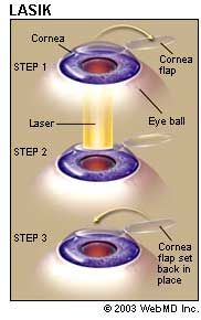
All laser vision correction surgeries work by reshaping the cornea, or clear front part of the eye, so that light traveling through it is properly focused onto the retina located in the back of the eye. LASIK is one of a number of different surgical techniques used to reshape the cornea.

What Are the Advantages of LASIK Eye Surgery?

LASIK has many benefits, including:
  • It works! It corrects vision. Around 80% of patients will have their desired vision after LASIK. An enhancement can further increase this number.
  • LASIK is associated with very little pain.
  • Vision is corrected nearly immediately or by the day after LASIK.
  • Usually no bandages or stitches are required after LASIK.
  • Adjustments can be made years after LASIK to further correct vision.
  • After having LASIK, most patients no longer need corrective eyewear.

What Are the Disadvantages of LASIK Eye Surgery?

Despite the pluses, there are some disadvantages to LASIK eye surgery:
lasik
  • Changes made to the cornea cannot be reversed after LASIK.
  • Corrections can only be made by additional LASIK.
  • LASIK is technically complex. Problems may occur when the doctor cuts the flap, which can permanently affect vision.
  • LASIK can cause a loss of "best" vision with or without glasses at one year after surgery. Your best vision is the highest degree of vision that you achieved while wearing your contacts or eyeglasses.

What Are the Potential Side Effects of LASIK Eye Surgery?

Some patients experience discomfort in the first 24-48 hours after LASIK eye surgery. Other side effects, although rare, may include:
  • Glare
  • Seeing halos around images
  • Difficulty driving at night
  • Fluctuating vision
  • Dry eyes

How Should I Prepare for LASIK Eye Surgery?

Before your LASIK eye surgery, you will meet with a coordinator who will discuss what you should expect during and after the surgery. During this session, your medical history will be evaluated and your eyes will be tested. Likely tests include measuring corneal thickness, refraction, corneal mapping, air pressure, and pupil dilation. Once you have gone through your evaluation, you will meet the surgeon, who will answer any questions you may have. Afterwards, you can schedule an appointment for the procedure.
If you wear rigid gas permeable contact lenses, you should not wear them for three weeks before your surgery. Other types of contact lenses shouldn't be worn for at least three days prior to surgery. Be sure to bring your eyeglasses to the surgeon so your prescription can be reviewed.
On the day of your surgery, eat a light meal before going to the doctor, and take all of your prescribed medications. Do not wear eye makeup or have any bulky accessories in your hair that will interfere with positioning your head under the laser. If you are not feeling well that morning, call the doctor's office to determine whether the procedure needs to be postponed.

What Happens During LASIK Eye Surgery?

During LASIK eye surgery, an instrument called a microkeratome is used to cut a thin flap in the cornea. The cornea is then peeled back and the underlying cornea tissue is reshaped using an excimer laser. After the cornea is reshaped so that it can properly focus light into the eye and onto the retina, the cornea flap is put back in place and the surgery is complete.
LASIK is performed while the patient is under a local anesthesia and usually takes about 10 minutes to complete.


What Should I Expect After LASIK Eye Surgery?

Healing after LASIK eye surgery usually occurs very rapidly. Most patients notice improved vision within a few days after surgery. However, your vision may be blurry and hazy for the first day. You should plan to have someone drive you home after LASIK.
Your eyes will be dry even though they do not feel that way. Your doctor will give you prescription eye drops to prevent infection and keep your eyes moist. These eye drops may cause a momentary slight burn or blurring of your vision when you use them. Do not use any eye drops not approved by your ophthalmologist. Specific follow-up after the surgery varies from one surgeon to another. You will revisit the doctor for an evaluation 24-48 hours after LASIK eye surgery, as well as at regular intervals within the first six months after surgery.


Read more »
These icons link to social bookmarking sites where readers can share and discover new web pages.
  • Digg
  • Sphinn
  • del.icio.us
  • Facebook
  • Mixx
  • Google
  • Furl
  • Reddit
  • Spurl
  • StumbleUpon
  • Technorati

Color blindness..


What is color blindness?

Color blindness means you have trouble seeing red, green, or blue or a mix of these colors. It’s rare that a person sees no color at all.
Color blindness is also called a color vision problem.

A color vision problem can change your life. It makes it harder to learn and read, and you may not be able to have certain careers. But children and adults with color vision problems can learn to make up for their problems seeing color.

What causes color blindness?

Most color vision problems are inherited (genetic) and are present at birth.
People usually have three types of cone cells in the eye. Each type senses either red, green, or blue light. You see color when your cone cells sense different amounts of these three basic colors. Most cone cells are found in the macula, which is the central part of the retina.
Inherited color blindness happens when you don't have one of these types of cone cells or they don't work right. You may not see one of these three basic colors, or you may see a different shade of that color or a different color. This type of color vision problem doesn't change over time.


A color vision problem is not always inherited. In some cases, a person can have an acquired color vision problem. This can be caused by:
  • Aging.
  • Eye problems, such as glaucoma, macular degeneration, cataracts, or diabetic retinopathy.
  • Injury to the eye.
  • Side effects of some medicines.

What are the symptoms?

The symptoms of color vision problems vary:
  • You may see many colors, so you may not know that you see color differently from others.
  • You may only be able to see a few shades of color, while most people can see thousands of colors.
  • In rare cases, you may see only black, white, and gray.

How is color blindness diagnosed?

Tests measure how well you recognize different colors.
  • In one type of test, you look at sets of colored dots and try to find a pattern in them, such as a letter or number. The patterns you see help your doctor determine which colors you have trouble with.
  • In another type of test, you arrange colored chips in order according to how similar the colors are. People with color vision problems cannot arrange the colored chips correctly.

How is it treated?

Inherited color vision problems cannot be treated or corrected.
Some acquired color vision problems can be treated, depending on the cause. For example, if a cataract is causing a problem with color vision, surgery to remove the cataract may restore normal color vision.

You can find ways to help make up for a color vision problem, such as wearing colored contact lenses or eyeglasses or wearing glasses that block glare. You can learn to look for other things, such as brightness or location, rather than colors. For example, you can learn the order of the three colored lights on a traffic signal.

How can you help a child who has color blindness?

Color vision problems can make learning and reading hard for children, which can lead to poor schoolwork and low self-esteem.
You can help your child by:
  • Making sure your child is tested for color vision problems during routine eye tests. The sooner you know there is a problem, the sooner you can help your child.
  • Telling your child’s teachers and other school staff about the problem. Suggest seating your child where there is no glare and using a color of chalk that your child can see.
Read more »
These icons link to social bookmarking sites where readers can share and discover new web pages.
  • Digg
  • Sphinn
  • del.icio.us
  • Facebook
  • Mixx
  • Google
  • Furl
  • Reddit
  • Spurl
  • StumbleUpon
  • Technorati

Stye..


What Is a Stye?

A stye is a pimple or abscess that forms in either the upper or lower eyelid. The medical term for stye is hordeolum (pronounced HOR-dee-oh-lumm) and it is an infection caused by bacteria that normally live on the surface of the eyelid skin without causing disease. Some of these germs get trapped along with dead skin cells inside crypts along the eyelid margin. Styes are usually superficial and plainly visible. Occasionally they can reside deeper within the eyelid.
An external stye starts as a pimple next to an eyelash. It turns into a red, painful swelling that may last several days before it bursts and then heals. Some external styes are short-lived and self-limiting while others may require the care of your eye doctor.
An internal stye (on the underside of the lid) also causes a red, painful swelling, but its location prevents the familiar whitehead from appearing on the eyelid. The internal stye may disappear completely once the infection is past, or it may leave a small fluid-filled cyst or nodule that can persist and may have to be opened and drained.
Folks tend to confuse a stye with another common lid lump, the chalazion (pronounced cha-LAY-zee-yon). A chalazion is very different from a stye and is not an infection. It is instead a firm, round, smooth, sometimes painless bump usually some distance from the edge of the lid. A chalazion is a local tissue reaction to oily glandular secretions that were unable to reach the lid surface because the duct was blocked by debris.
Styes and chalazia are usually harmless and rarely affect your eyeball or your eyesight but in some cases they can lead to severe infections of the face called cellulitis which can be very serious. They can occur at any age and tend to periodically recur.

What Causes Styes?

Styes are usually caused by staphylococcal bacteria, which often live right on the skin surface. Our bodies are coated with billions of friendly bacteria that coexist with us. When the conditions are just right the bacteria feast on dead cells and other debris, resulting in the tender pimple.
A chalazion is caused by blockage of tiny ducts in the eyelid glands that normally transport an oily substance. This oily material (sebum) is the layer of the tear film that reduces tear evaporation. Trapped or misplaced oil stimulates the immune system to clean up the mess. Chalazia develop over weeks to months.
Read more »
These icons link to social bookmarking sites where readers can share and discover new web pages.
  • Digg
  • Sphinn
  • del.icio.us
  • Facebook
  • Mixx
  • Google
  • Furl
  • Reddit
  • Spurl
  • StumbleUpon
  • Technorati